
March 9, 2020 - Trying out New Color Segmentation Method Part 2
Lab Work
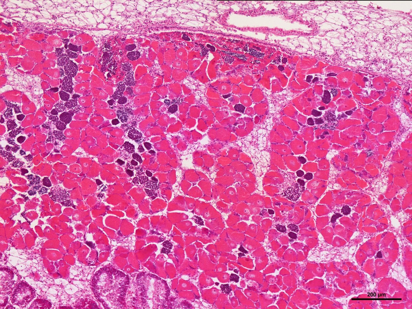
Last week, I began to try and find a different way to segment the histology images by color in order to find a faster and possibly more accurate way to threshold the images instead of the method that I was previously using. The new way that I found which might be useful is seen in the article “Color spaces in OpenCV (C++ / Python)”, and I began to follow the steps used in the article to segment images by color on this image:
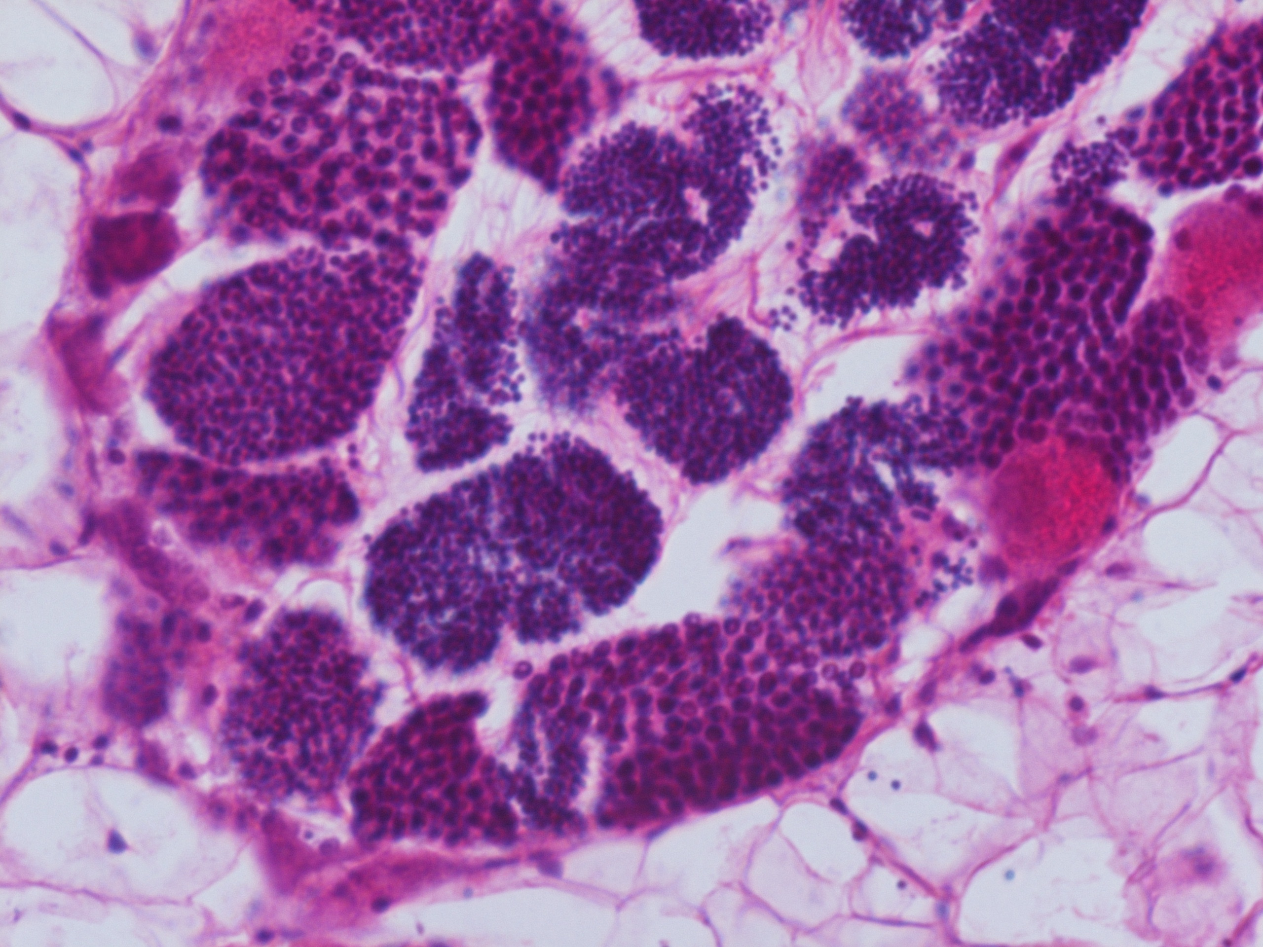
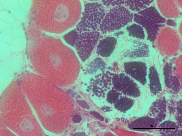
But after looking at the article a little more, I realized that they were using mutiple images in order to compare what the color spaces look like for the same object under different lightings. Thus, I used two more images (seen below) to use as comparisons in this process:
I compared the 3 images through 4 color spaces: RGB, Lab, YCrCb, and HSV. The results are shown below:
original: 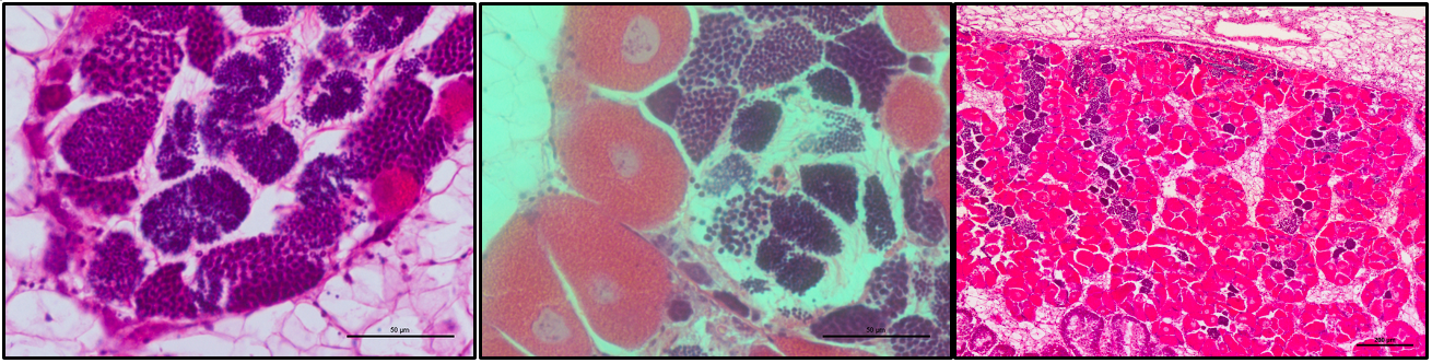 R:
R: 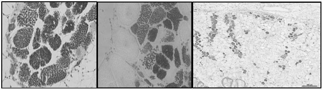 G:
G: 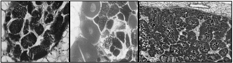 B:
B: 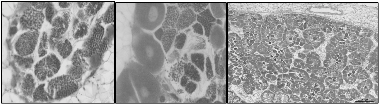 L:
L: 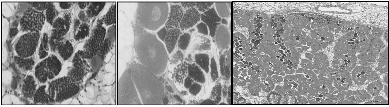 a:
a: 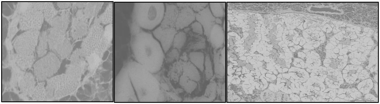 b:
b: _comparison.png?raw=true) Y:
Y: 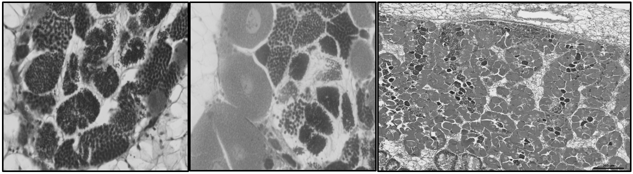 Cr:
Cr: 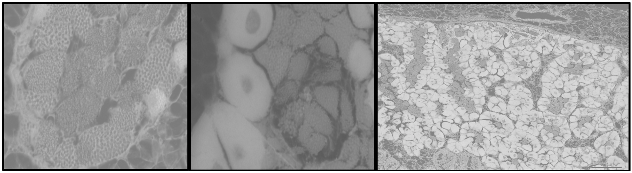 Cb:
Cb: 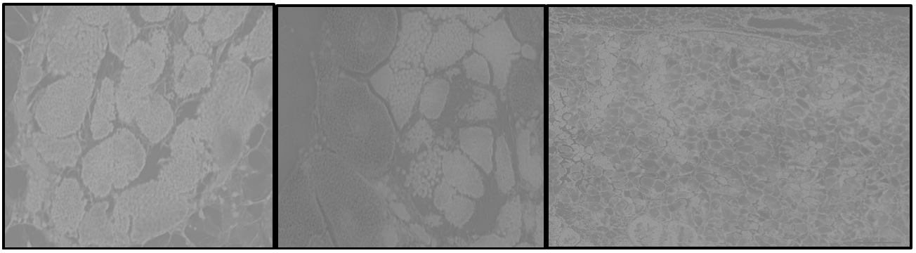 H:
H: 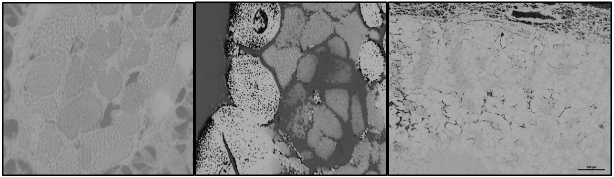 S:
S: 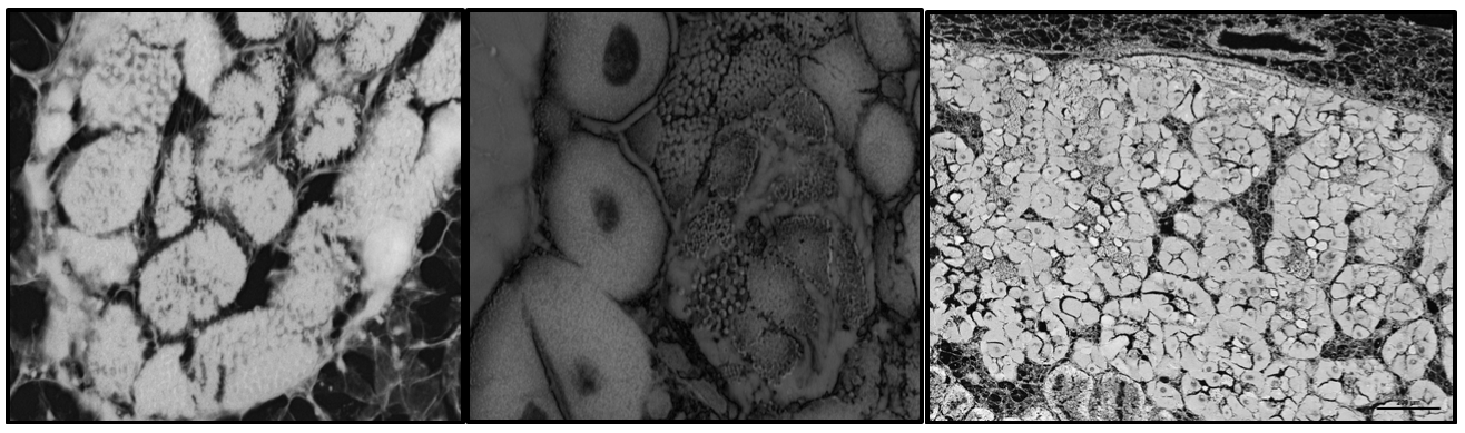 V:
V: 
Next Steps
Compare the color spaces of these images (finding similarities and differences) and continue working through the article tutorial